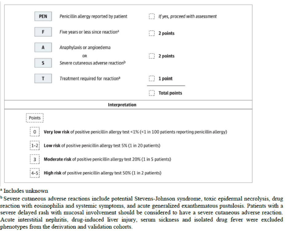
Case 1: Influenza – Stacey

Stacey, a 60-year-old woman, comes into your clinic with muscle aches, fever, and general fatigue, which came on suddenly three days ago. Previously, she had felt well. She is not in distress and her vital signs are normal.

Based on her history and a physical examination, you feel there is a high likelihood she has influenza or another viral, flu-like illness. A rapid test for influenza A is positive, in the clinic. Stacey states that she would like antibiotics in order to feel better. 

How would you approach your discussion with the patient?

Your response to Stacey can explain that antibiotics will not help her feel better. Antibiotics are not indicated, nor expected to be of benefit for influenza. Influenza is caused by a virus and antibiotics cannot cure a virus. It would be appropriate to discuss with her that the natural history of influenza infection, or what we normally expect in the case of flu, would suggest that her symptoms are likely to improve within 5-7 days. You can also give her clear information about when antibiotics would be beneficial. You can advise that she should return if she develops symptoms that point towards a secondary bacterial infection, such as persistent fever, increasing difficulty breathing, or if her symptoms do not improve over the next 3-4 days.

A key component in this case is effectively communicating with the patient to ensure they understand the reasoning behind not prescribing antibiotics. Often, providing a written handout can ensure that your patient retains the key messages you have discussed and may help them feel they have ‘gotten something’ out of the appointment. One such resource is the Viral Prescription Pad (see References). This resource helps patients understand their diagnosis, gives recommended over-the-counter analgesics they can use, and instructions to return if certain symptoms arise.

This would also be an ideal time to discuss the benefits of the annual influenza vaccination and health promotion strategies for the future. You can remind Stacey that building good hand washing habits is a simple and effective way to prevent the flu.  

Sometimes good antimicrobial stewardship practice involves knowing when medications other than antibacterial agents are likely to be of benefit. It is important to note, although not applicable in this case, that there are certain factors that put patients with influenza virus infection at a higher risk for progressive disease. These include severe disease (requiring hospitalization); however, some patients with milder disease are at risk for severe disease (see Risk Factors). In these patients with suspected or proven influenza, antiviral medication (oseltamivir) would be indicated if they presented to clinic within 48 hours of symptom onset to reduce the risk of severe outcomes such as hospitalization or death. 


Risk Factors:

Major risk factors that predispose individuals to severe influenza (Adapted from Aoki et al, 2019)

  • Severe chronic illness, including asthma and other chronic pulmonary diseases, cardiovascular disease, chronic renal insufficiency, diabetes mellitus, and chronic neurological or neudevelopmental disorders
  • Obesity with BMI ≥40
  • Age younger than 5, or older than 64
  • Immunosuppression due to disease (eg. HIV infection with CD4 is < 200 × 106/L) or medication
  • Pregnant women and women up to 4 weeks postpartum

References: 

Case 2: Community-Acquired Pneumonia – Jesse

Jesse, a 5-year-old boy, presents to your office with fever, cough, and tachypnea. These symptoms started 3 days ago. He has been eating well throughout this time and has stable vital signs when the pediatrician examines him. 

A rapid test, performed by the clinic nurse, is negative for SARS-CoV2 virus. The chest X-ray results are shown below: 

X-ray image of lungs with an infection.
Image Credit: https://www.intechopen.com/chapters/42153 (Accessed Sept 17, 2022).

Based on the clinical symptoms and chest X-ray findings, you determine that Jesse is well enough to be treated as an outpatient, but will still require antibiotics to help clear the infection. 

Given the circumstances of this case, which antibiotic would you prescribe for Jesse? 

  1. Clindamycin 
  2. Amoxicillin-Clavulanic acid (Amox-Clav)
  3. Amoxicillin
  4. Azithromycin

The best choice in this case is (c) amoxicillin. The findings on chest X-ray indicate a bacterial etiology of the community-acquired pneumonia (CAP), therefore, it is appropriate to treat with antibiotics. Streptococcus pneumonia is the most common bacterial cause of CAP and knowing this will help with the choice of antibiotic.Amoxicillin is a very effective antibiotic against S. pneumoniae and is recommended to treat community acquired bacterial pneumonia by the Canadian Paediatric Society.

An important part of preventing antimicrobial resistance is choosing the most effective and narrow spectrum antibiotic for the situation. Antibiotics should be chosen to be the narrowest spectrum possible while still targeting the causative bacteria, which in this case would be amoxicillin. The other antibiotic options in this case are broader spectrum antibiotics, but would not be appropriate for this situation. Broad spectrum antibiotics can have a greater effect on the normal flora, or microbiome, of the patient. This can contribute to antimicrobial resistance by allowing overgrowth of drug-resistant bacteria within the microbiome of the patient. 

In this case, CAP was caused by bacteria, but bacteria are actually the second most common cause of CAP. The most common pathogens responsible for CAP in healthy children are respiratory viruses, especially during the winter. Some common viral causes include respiratory syncytial virus, influenza virus, parainfluenza virus and human metapneumovirus. 

The duration of treatment for children with uncomplicated community acquired pneumonia who are well enough to be treated as outpatients is also an important question. In adults, 5-7 days has been shown to be as effective as longer courses; in children, the typical antibiotic course has been somewhat longer (7 to 10 days) by historical practice. However, a recent RCT demonstrated that, similar to adults, in children aged 6 months to 5 years old with non-severe CAP, 5 days of antibiotic therapy is as effective as 10 days (see References). Practice guidelines are increasingly adopting the shorter, equally effective duration of 5 days.


References: 

  • Le Saux N, Robinson JL. Uncomplicated pneumonia in healthy Canadian children and youth: Practice points for management. Paediatr Child Health. 2015 Nov-Dec;20(8):441-50. doi: 10.1093/pch/20.8.441.
  • Canadian Pediatric Society 2018 “Uncomplicated pneumonia in healthy Canadian children and youth: Practice points for management”. See:  https://cps.ca/en/documents/position/pneumonia-management-children-youth
  • Greenberg D, Givon-Lavi N, et al. Short-course antibiotic treatment for community-acquired alveolar pneumonia in ambulatory children: a double-blind, randomized, placebo-controlled trial. Pediatr Infect Dis J 2014. Feb;33(2):136-42. doi: 10.1097/INF.0000000000000023.

Case 3: Urinary Tract Infection – Ming

Ming is a 78-year-old woman with cognitive impairment and living in a long-term care facility. She is seen by you, her primary care practitioner, after the nurse taking care of her over the weekend called for foul smelling urine. On review, she denies any other symptoms and her nursing team affirms that she is in her usual state of health and her vital signs are normal. A mid-stream urine sample is taken for analysis and is noted to be dark yellow and cloudy. Urine dipstick testing reveals a specific gravity (S.G.) of 1.030, a pH of 6.6, trace protein, a positive leucocyte esterase and nitrite test, and negative results for blood, glucose, and ketones. Over the weekend, the team overseeing her care suspected she might have a UTI, so a urine culture was sent to the microbiology lab. Today, the lab reports a positive test result with >10^8 colony-forming units per L of E. coli, which is significant. 

What do you do next?

  1. Treat with nitrofurantoin for 5 days 
  2. Treat with ciprofloxacin for 3 days 
  3. Treat with trimethoprim/sulfamethoxazole for 5 days
  4. No treatment necessary
  5. Repeat the urine culture

BEST ANSWER: 4. No treatment necessary

Teaching Points:

Asymptomatic bacteriuria (ASB) is the presence of one or more species of bacteria growing in the urine (≥10^5 colony-forming units [CFU]/mL or ≥10^8 CFU/L), regardless of the presence of pyuria, in the absence of signs or symptoms attributable to urinary tract infection (UTI). The patient described here does not have any signs or symptoms that are associated with UTIs, which include dysuria and urinary frequency, (see UTI Symptoms). Cloudy or foul-smelling urine are not indicative of a urinary tract infection in the absence of symptoms.

A positive urine leucocyte esterase test reliably identifies pyuria but may be seen in other inflammatory disorders of the pelvis or genitourinary tract. A nitrite test is also of limited value because urinary tract infections can still occur with non-nitrite producing uropathogens. False positives can also be seen with delays in sample testing. Although the combination of a positive leucocyte esterase and a positive nitrite is more specific to bacteriuria, these tests do not distinguish between those patients with asymptomatic bacteriuria and those that are symptomatic (i.e. have a UTI). 

To summarize, a urinalysis that is negative for both nitrites and pyuria does indicate that a UTI is unlikely, but even a positive urinalysis for both nitrites and pyuria in absence of symptoms (see Box) does not ‘rule in’ a UTI, and could indicate ASB.

ASB should not be treated. Exceptions to this rule include treatment of ASB in pregnant women and prior to an invasive urological procedure where bleeding can occur. Further, changes to urine such as foul-smelling or cloudy urine, in the absence of symptoms of a UTI, should not prompt testing. A urine culture in an asymptomatic patient (excluding those with indications listed above) is unnecessary and can lead to wasted healthcare resources, as well as unnecessary antibiotic prescriptions. Antibiotic therapy exposes patients to risk of adverse effects and promotes antibiotic resistance. Caution against unnecessary treatment should be taken, especially for older adults living in long-term care homes where the prevalence of ASB can be up to 50%. 


UTI Symptoms & Signs:

Cystitis                       Dysuria

                                    Urinary frequency

                                    Hematuria

                                    Lower abdominal/suprapubic pain

                                    Dyspareunia

Pyelonephritis*         Fever

                                    Chills/rigors

                                    Flank pain (or costovertebral angle tenderness)

*Patients with pyelonephritis can also present with symptoms of cystitis


Resources: 

  • Rx Files: A trusted Canadian resource with general information on asymptomatic bacteriuria. Updated periodically. Available here.
  • StatPearls: Further reading and high yield tips on ASB, updated regularly. Available here.
  • Guidelines: The Infectious Diseases Society of America published guidelines on the management of asymptomatic bacteria in 2019. See Clinical Infectious Diseases, available here
  • Toolkits: AMMI Canada has an antibiotic stewardship campaign called ‘Symptom Free Pee’, with tools to support an appropriate approach to ASB and UTIs in the elderly, including a ‘myths and truths’ FAQ, posters, and other resources for use in acute and long-term care settings. Available here.

Case 4: Urinary Tract Infection – Mary-Elle

Mary-Elle is a 25-year-old woman who comes to see you in your office. She describes a two-day history of dysuria, urgency, and frequency, but denies flank pain. She is afebrile, with no costovertebral angle tenderness. She has had a urinary tract infection previously, the most recent being over a year ago, and recognizes her current symptoms as very typical for her. A urine dipstick urinalysis is done in your office, which reveals a specific gravity (S.G.) of 1.030, a pH of 6.2, no proteins, a positive leucocyte esterase and nitrite test, and a positive result for blood but negative results for glucose and ketones. You suspect that Mary-Elle has a UTI.

What do you do next?

  1. Treat with nitrofurantoin for 5 days
  2. Treat with ciprofloxacin for 3 days
  3. Confirm they have a UTI with a urine culture, and wait for the results before treating
  4. Send for a urine culture, and treat with ciprofloxacin for 3 days
  5. No treatment necessary

BEST ANSWER: 1. Treat with nitrofurantoin for 5 days

Teaching Points:

This patient has cystitis, an infection of the lower urinary tract which causes dysuria, with or without frequency, urgency, and suprapubic pain (see UTI Symptoms). The presence of fever or costovertebral pain or tenderness, which is absent here, would alert you to the possibility of pyelonephritis. In women without urinary catheters or structural urologic abnormalities, cystitis (i.e. uncomplicated lower UTI) is a clinical diagnosis. Confirmation with urinalysis in the office is, however, reasonable, given the high sensitivity of the test for the presence of bacteriuria and UTI. The absence of both pyuria and nitrites can help rule out UTI, especially if the patient’s symptoms are slightly atypical. Routine urine culture is not recommended for women who present with clear symptoms of uncomplicated cystitis.

UTI is a clinical diagnosis and should be treated empirically. Treatment decisions should always be verified by up-to-date guidelines, while considering local antibiotic resistance patterns. First-line options include a 5-day course of nitrofurantoin or a 3-day course of TMP-SMX. Ciprofloxacin may be a reasonable second-line treatment option but is best avoided as a first-line choice due to the overly broad spectrum, which increases the risk of promoting the development of drug-resistant organisms, as well as the risk for C. difficile.

Certain exceptions should be noted. If the patient’s symptoms were somewhat atypical, it would be reasonable to test for a UTI with a urine culture. Other reasons to consider urine culture would be a history of culturing antimicrobial resistant bacteria, or in patients with a history of recent or recurrent UTI . However, this is not the case here, and the pre-test probability prior to urine culture is already high enough to treat.


UTI Symptoms & Signs

Cystitis                       Dysuria

                                    Urinary frequency

                                    Hematuria

                                    Lower abdominal/suprapubic pain

                                    Dyspareunia

Pyelonephritis*          Fever

                                    Chills/rigors

                                    Flank pain (or costovertebral angle tenderness)

*Patients with pyelonephritis can also present with symptoms of cystitis


Resources:

  • Bugs & Drugs App:  A free, regularly updated, Canadian reference for appropriate use of antimicrobials. Contains recommendations for treatment of UTI, including cystitis. Available at the App Store or Google Play.
  • Guidelines: The Infectious Diseases Society of America published guidelines on the management of uncomplicated urinary tract infections in 2011. These guidelines are currently under review. Clinical Infectious Diseases, Volume 52, Issue 5, 1 March 2011, Pages e103–e120. Available here.  

Case 5: Urinary Tract Infection – Tharmala

Tharmala is a 30-year-old woman who is 11 weeks pregnant and presents for her first prenatal appointment. She is in good general health and denies any symptoms specific to the urinary tract. As part of the general screening, you request a urine culture. The results come back with >10^8 CFU/L of E. coli, which you know is significant. Antimicrobial susceptibility testing for the E. coli shows it to be susceptible to Trimethoprim-sulfamethoxazole, fluoroquinolones, amoxicillin, and cephalexin, but resistant to nitrofurantoin.

What do you do next?

  1. Treat with Trimethoprim-sulfamethoxazole for 5 days
  2. Treat with ciprofloxacin for 3 days
  3. Treat with amoxicillin for 5 days
  4. Treat with nitrofurantoin for 5 days
  5. No treatment necessary

BEST ANSWER: 3. Treat with amoxicillin for 5 days

Teaching Points:

Based on guidelines, asymptomatic bacteriuria (ASB) is defined as >10^8 CFU/L of uropathogens identified on urine culture, which is still present on repeat urine culture testing. Repeat testing will help to exclude the possibility that your patient is among the ~20% of individuals who will spontaneously clear the ASB. Practically speaking, however, a repeat urine culture is rarely pursued in clinical practice.

In most patients, asymptomatic bacteriuria should not be treated, but in pregnant women, screening for ASB during the first trimester, or on their first obstetrical visit, is indicated. If the screening urine culture is positive, treatment is indicated to reduce the risk of pyelonephritis and spontaneous abortion. Pregnant individuals are tested routinely during pregnancy and, if urine cultures are positive for uropathogens, are treated.

Beta-lactam antibiotics such as amoxicillin or cephalexin are the safest choices of antibiotics to give to someone during pregnancy. Nitrofurantoin is also a safe choice in pregnancy, except in patients at term (i.e. 38-42 weeks), due to the risk of hemolytic anemia in the newborn.  However, nitrofurantoin is noted to be resistant in this case. Trimethoprim-sulfamethoxazole is contraindicated in the first trimester due to its action as a folate antagonist causing neural tube defects. Also, fluoroquinolones are generally not recommended during pregnancy due to potential fetal bone toxicity.


Resources:

  • Guidelines, Screening: The Canadian Task Force on Preventative Healthcare published guidelines on screening for ASB in pregnancy, which can be found here:
  • Guidelines, Clinical Management: The Infectious Diseases Society of America has also published guidelines on the management of asymptomatic bacteria, in 2019. See Clinical Infectious Diseases,  Available here.
  • Bugs & Drugs App: A Canadian app that is regularly updated, downloadable, and compatible with iPhone and Android devices. Available at the App Store or Google Play.

Case 6: Community-Acquired Pneumonia – Doug

Doug, a 67-year-old man, is brought into the emergency room by his partner who is concerned that he is acting confused and disoriented. Doug can provide minimal history, but his partner, Brad, reports that he has been feeling unwell for the past week, with a productive cough, shortness of breath, and maybe a fever.

Doug has not recently been hospitalized or had any infections. Brad can’t remember the last time Doug had to use antibiotics. They have not traveled recently, and apart from their dog, Ralph, Brad recalls no recent animal exposures.

On exam, Doug has a temperature of 36.9°C, HR 128 bpm, RR 25 bpm, BP 110/70, O2 saturation 89% on room air, with warm and flushed skin. He appears confused, with a decreased level of consciousness (Glasgow Coma Scale of 12/15). On respiratory exam, you hear decreased breath sounds on the right side as well as dullness on percussion over the right lower lobe. 

Initial bloodwork shows leukocytosis with a white blood cell count (WBC) of 13.1 x 10^9/L, hemoglobin 160 g/L, platelets 550 x 10^9/L, a lactate level of 4.0mmol/L, a serum creatinine level of 150 umol/L, sodium 140mmol/L, potassium 4.9mmol/L, chloride 98mmol/L. ALT 100U/L, AST 95 U/L, ALP 168U/L. His VBG shows a pH 7.32, PCO2 30mmHg, P02 55mmHg, and a HC03 15mmol/L. Blood cultures are taken from two peripheral sites. You order a chest x-ray, and the results are shown below.
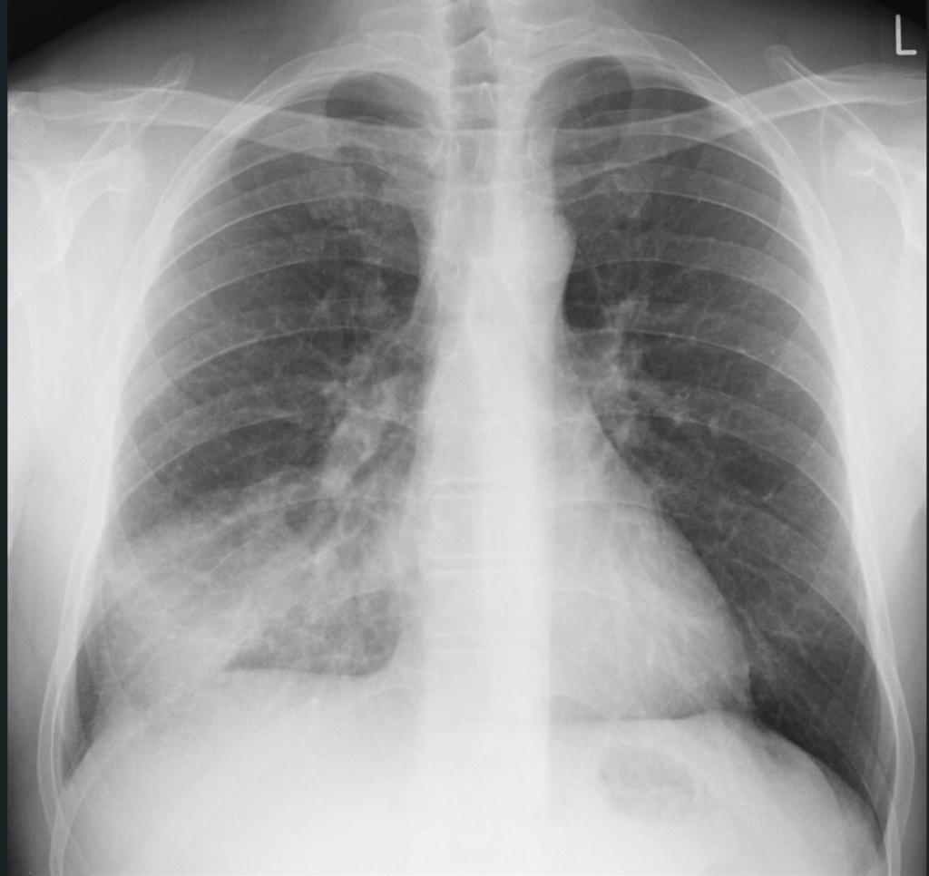
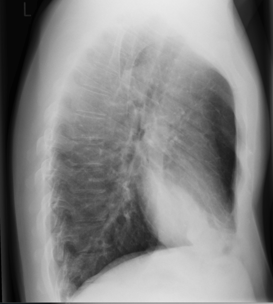
Radiographic images courtesy of Sajoscha A. Sorrentino, Radiopaedia.org, rID: 14979

The x-ray reveals an infiltrate in the right middle and lower lobe, with no effusion of radiographic evidence of heart failure.

Question 1: In addition to community-acquired pneumonia, what is the most appropriate diagnosis?

  1. Pre-sepsis
  2. Systemic inflammatory response syndrome
  3. Sepsis
  4. Severe sepsis
  5. Septic shock

BEST ANSWER: C. Sepsis

Teaching Points:

Sepsis is defined as life-threatening organ dysfunction caused by a dysregulated host response to infection. The patient in this scenario is septic, as shown by his decreased level of consciousness, hypoxia, acute kidney injury, and elevated lactate in the context of suspected community acquired pneumonia. A bedside clinical assessment, with or without a combination of screening tools (SIRS, qSOFA, MEWS, NEWS), in addition to a few basic laboratory tests, including lactate (>2 mmol/L), can help to determine if a patient has or is at risk for sepsis. Doug did not present with a fever, but this may be absent, especially on initial assessment. Fever or leukocytosis are often present, but neither are sensitive enough to rule out a diagnosis of sepsis.

At the bedside, you can recognize a patient with a suspected or proven infection with sepsis if they are showing evidence of organ dysfunction due to this infection (see Definitions and Bedside Clinical Criteria). Septic shock defines a more severe subset of patients with sepsis. Patients with septic shock require vasoactive medications to support their blood pressure, even after adequate fluid resuscitation. Systemic inflammatory response syndrome and severe sepsis are no longer used to identify sepsis.

The diagnosis of sepsis and septic shock helps to determine the prognosis for the patient, as well as the initial urgency of management. Sepsis is associated with an in-hospital mortality of approximately 10% versus 30% for septic shock.


Definitions and Bedside Clinical Criteria

SepsisSeptic Shock
DefinitionLife-threatening organ dysfunction due to a dysregulated physiologic response to infection.Sepsis with profoundly abnormal circulatory, cellular, and metabolic changes. Greater risk for mortality compared to sepsis alone.
Bedside Criteria to Look forEvidence of infection, plus major organ dysfunction. This can be measured using a “SOFA*” score of 2 or more.Hypotension requiring vasopressor medications to maintain blood pressure, and lactic acidosis, despite fluid resuscitation.

*SOFA, the Sequential Organ Failure Assessment Score, uses clinical evidence of organ dysfunction, including low oxygenation, blood pressure, level of consciousness, and platelet counts, as well as elevated bilirubin and creatinine, to provide a score between 0 and 24. Higher scores are correlated with worse prognosis. See the online calculator here.


Question 2: What are the possible courses of action to treat the infection?

Course of Action

  1. Obtain blood cultures from two peripheral sites, as well as a nasal culture for MRSA*, and wait for results before starting antibiotics. Start targeted antibiotics based on susceptibility (for example, ampicillin for penicillin-susceptible Streptococcus pneumoniae).
  2. Obtain blood cultures from two peripheral sites, as well as nasal culture for MRSA*, and treat empirically with piperacillin-tazobactam and vancomycin for five days. There is no need to narrow the antibiotics while he is in hospital.
  3. Treat empirically with piperacillin-tazobactam and vancomycin stat and continue for five days. There is no need to obtain blood cultures or narrow the antibiotics while he is in hospital.
  4. Begin ceftriaxone, azithromycin and vancomycin stat, and then obtain blood cultures from two peripheral sites, as well as nasal culture for MRSA*. Narrow antibiotics based on susceptibility.
  5. Obtain blood cultures from two peripheral sites and nasal culture for MRSA*, then begin ceftriaxone, azithromycin and vancomycin stat. Narrow antibiotics in 48-72 hours based on clinical response and susceptibility, once available.
    • *Methicillin-resistant Staphylococcus aureus

Resources:

  • Bugs & Drugs App: A free, regularly updated, Canadian reference for appropriate use of antimicrobials. Contains recommendations for treatment of community-acquired pneumonia, including in patients with sepsis (i.e. patients requiring hospitalization). Available at the App Store or Google Play.
  • Guideline, CAP: Metlay JP, Waterer GW, Long AC, et al. Diagnosis and treatment of adults with community-acquired pneumonia. An official clinical practice guideline of the American Thoracic Society and Infectious Diseases Society of America. Am J Respir Crit Care Med. 2019;200(7):e45-e67. doi:10.1164/rccm.201908-1581ST
  • Guideline, Sepsis & Septic Shock: Rhodes A, Evans LE, Alhazzani W, et al. Surviving sepsis campaign: international guidelines for management of sepsis and septic shock: 2021 update. Crit Care Med. 2021;49(3):e1063-e1143. doi:10.1097/CCM.0000000000004899
  • Sepsis & Septic Shock Definitions: Singer M, Deutschman CS, Seymour CW, et al. The Third International Consensus Definitions for Sepsis and Septic Shock (Sepsis-3). JAMA. 2016;315(8):801-810. doi:10.1001/jama.2016.0287
  • Sequential Organ Failure Assessment (SOFA) Score: An online calculator of the SOFA Score is available here.

Teaching Points: 

Once identified, antimicrobials should be administered ‘stat’ to a patient with suspected sepsis, after blood cultures are drawn. In patients with septic shock, antibiotics should be given within one hour of recognition. Timely appropriate antibiotic therapy has been associated with a reduction in mortality. Sputum cultures are also recommended for patients with suspected sepsis due to pneumonia, but antibiotics should not be delayed while awaiting their collection.

In this patient with suspected sepsis and community-acquired pneumonia, common bacterial pathogens include S. pneumoniae, H. influenzae, Legionella pneumophila, and S. aureus. Broad spectrum antibiotics are used initially, with regular assessment for de-escalation of antimicrobials. Local guidelines, which incorporate the prevalence of drug resistant bacteria (including MRSA), should be consulted for specific empiric antimicrobial choices. The 2019 IDSA/ATS guidelines on the Diagnosis and Treatment of Community Acquired Pneumonia recommend treating patients with severe community-acquired pneumonia (i.e. those that have sepsis) with a combination of a 3rd generation cephalosporin (such as cerftriaxone) and either a macrolide (such as azithromycin) or a respiratory fluoroquinolone (such as levofloxacin). Vancomycin should be considered if MRSA is highly prevalent in the community. If vancomycin is added, MRSA colonization testing of the nares should be performed to allow for de-escalation (stopping) vancomycin at 48-72 hours, assuming nasal and blood cultures are negative for MRSA.

Most blood cultures for pathogenic organisms, such as S. pneumoniae, will be positive within 48 hours, thus, 48-72 hours is an appropriate time to reassess antimicrobials that were started empirically for a septic patient such as this one. The administration of antimicrobials before blood cultures are taken can significantly reduce the yield of blood cultures. Therefore, it is important to make every effort to collect blood cultures before the administration of systemic antibiotics in patients with suspected sepsis.

Fixed durations of therapy without regular reassessment for de-escalation, as proposed in choices b. and c., is not recommended. Furthermore, while piperacillin-tazobactam and vancomycin provide good coverage of S. pneumoniae, H. influenzae, and S. aureus, these antimicrobials would not be active against Legionella and the other “atypical” bacterial pathogens that can cause community-acquired pneumonia.  The added Gram-negative coverage provided by pipericillin-tazobactam (as opposed to a 3rd generation cephalosporin) would provide minimal benefit and is unnecessary here.

Case 7: C. diff – Margaret

Margaret is a 74-year-old patient who underwent a successful left knee replacement surgery and is recovering well. She has a past medical history of: Osteoarthritis, chronic kidney disease, COPD, gout, GERD. She has a recent history of Clostridioides difficile infections, the first of which occurred after a course of levofloxacin prescribed for pneumonia 2 years ago. She was treated with a 10-day course of oral Vancomycin and recovered completely. Unfortunately, she developed a C. difficile infection again, this time in hospital after her recent left knee replacement, once again successfully treated with a 14-day course of Vancomycin.
Margaret is seen in follow-up. She is planning for an elective right knee replacement (the other knee) in the next 3 months. She is worried about recurrence and, as part of perioperative planning, a repeat C. difficile sample was obtained. This revealed a positive result for C. difficile in her stool. She is not currently experiencing diarrhea and reports one bowel movement a day, which is usual for her.

In preparation for her surgery, what would you recommend for treatment of her C. difficile?

  1. No treatment is necessary.
  2. Vancomycin 125 mg PO QID for 10 – 14 days
  3. Vancomycin 125 mg PO QID for 10 – 14 days; followed by a ‘taper and pulse’ extended regimen of vancomycin (e.g., 125 mg PO TID for 7 days, 125 mg PO BID for 7 days, 125 mg PO once daily for 7 days, and then every 3 days for 2 – 3 weeks)
  4. Fidoxamicin 100 mg PO BID for 10 days
  5. Metronidazole 500 mg PO TID for 10 – 14 days

BEST ANSWER: A. No treatment necessary

  1. No treatment is necessary.
  2. Vancomycin 125 mg PO QID for 10 – 14 days
  3. Vancomycin 125 mg PO QID for 10 – 14 days; followed by a ‘taper and pulse’ extended regimen of vancomycin (e.g., 125 mg PO TID for 7 days, 125 mg PO BID for 7 days, 125 mg PO once daily for 7 days, and then every 3 days for 2 – 3 weeks)
  4. Fidoxamicin 100 mg PO BID for 10 days
  5. Metronidazole 500 mg PO TID for 10 – 14 days

Teaching Points: Clostridioides difficile is a common cause of antibiotic-associated colitis. Transmitted via the fecal-oral route, colonization of the intestinal tract with C. difficile also occurs in 3-5% of healthy adults. Risk factors for disease due to C. difficile (termed C. difficile infection, CDI) include antibiotic use, which is the most common and modifiable factor, as well as older age, hospitalization, and chronic comorbid illness.

Diagnosis of C. difficile infection is based on the presence of clinical symptoms (unexplained, new onset diarrhea of ≥ 3 unformed stools in 24 hours) along with a diagnostic test that detects C. difficile organism or toxin production. In Manitoba, C. difficile testing is performed using a 2-stage approach, initially with GDH antigen screen (very sensitive, poor specificity) followed by testing for C. difficile toxin A & B via nucleic acid amplification testing (NAAT). It is important to recognize that, in some cases, both recent treatment of C. difficile, as well as asymptomatic carriage of C. difficile can lead to false positive results. This can lead to over treatment. For this reason, in accredited clinical microbiology laboratories, requests for C. difficile assay on fully formed stool will be rejected by the lab (and, probably should have been rejected in this case).

Margaret does have a history of two previous C. difficile infections that required treatment and is at risk for recurrence.  Although her repeat C. difficile toxin test came back positive, it’s important to consider the clinical context; she is asymptomatic, and her positive result may be due to recent treatment of C. difficile infection, rather than an active (current) disease, and requires no treatment. More importantly, in view of her upcoming surgery, in the context of recent C. difficile, extra care should be taken to avoid unnecessary antimicrobial pressure, which could result in a significant risk of recurrent C. difficile. Strategies to reduce her risk of recurrent C. difficile after her next surgery include avoiding unnecessary antibiotics (for example, keeping perioperative antibiotic prophylaxis to the shortest possible course), as well as not testing for, or treating, asymptomatic bacteriuria if it develops post-op.

For information, see first line treatment options (Box, inset) for C. difficile (i.e., in persons who are symptomatic).


First Line Treatment Options

Box: First line treatment for patients with (symptomatic) non-fulminant* C. difficile infection

  • Stop systemic antimicrobials for other indications, if possible
  • Vancomycin 125 mg PO QID for 10 to 14 days
  • Alternative: fidaxomicin 200 mg PO BID for 10 days**
  • Combination therapy (such as with vancomycin and metronidazole) is not recommended

*Patients with fulminant (also known as severe-complicated) C. difficile have shock, persistent hypotension, ileus or toxic megacolon.

**Vancomycin and fidaxomicin are equally effective at curing mild, moderate, or severe (but non-fulminant) C. difficile. Fidaxomicin is associated with a slightly lower risk of relapse (number needed to treat to prevent one relapse of 9-10 patients), but due to increased cost of this agent and drug coverage, it is often not used as first line.

Metronidazole 500 mg PO TID, which is inferior in patients with severe C. difficile, is a safe and inexpensive option, which may also be used as an alternative in patients with non-severe C. difficile (including those with normal white blood cell count & no acute kidney injury).


References:

  • Loo VG, Davis I, Embil J, Evans GA, Hota S, Lee C, et al. (2018). Association of Medical Microbiology and Infectious Disease Canada treatment practice guidelines for Clostridium difficile infection. Journal of the Association of Medical Microbiology and Infectious Disease Canada, 3(2), 71-92. doi.org/10.3138/jammi.2018.02.13. Available here.
  • Johnson S, Lavergne V, Skinner AM, et al. Clinical Practice Guideline by the Infectious Diseases Society of America (IDSA) and Society for Healthcare Epidemiology of America (SHEA): 2021 Focused Update Guidelines on Management of Clostridioides difficile Infection in Adults. Clin Infect Dis. 2021;73(5):755-757. doi:10.1093/cid/ciab718. Available here.
  • Kelly CR, Fischer M, Allegretti JR, et al. ACG Clinical Guidelines: Prevention, Diagnosis, and Treatment of Clostridioides difficile Infections [published correction appears in Am J Gastroenterol. 2022 Feb 1;117(2):358]. Am J Gastroenterol. 2021;116(6):1124-1147. doi:10.14309/ajg.0000000000001278. Available here.

Case 8: Skin Abscess – Cheryl

Cheryl, a 46-year-old female, presents to her primary care clinic with a new carbuncle on her upper back that developed over the last 4 days. She tells you that it is quite painful, but does not report any fevers, chills, or night sweats. She has a past medical history of type 2 diabetes and hypertension; a recent culture of a previous foot ulcer 2 months prior was positive for growth of methicillin-resistant Staphylococcus aureus (MRSA). Her regular medications are metformin, Sitagliptin and ramipril. Upon examination, she looks well otherwise, with a fluctuant, erythematous subcutaneous nodule presenting on the skin above her left scapula which measures approximately 3 cm of induration and erythema in diameter.

Cheryl asks you what would be the next best course of action?

  1. Incision and drainage (I&D) with a 7-day prescription for TMP/SMX
  2. A 7-day course of Cephalexin  
  3. A 7-day prescription for Doxycycline and Cephalexin
  4. I & D, with a prescription for 10 days of Cephalexin and Doxycycline
  5. I & D, with no antibiotic prescription at this time

BEST ANSWER: E. I & D, with no antibiotic prescription at this time

  1. Incision and drainage (I&D) with a 7-day prescription for TMP/SMX
  2. A 7-day course of Cephalexin  
  3. A 7-day prescription for Doxycycline and Cephalexin
  4. I & D, with a prescription for 10 days of Cephalexin and Doxycycline
  5. I & D, with no antibiotic prescription at this time

Teaching point: Incision and drainage is indicated as first line management for all patients with simple or uncomplicated abscesses and boils, to help fascilitate drainage of purulent material, relieve pain, and promote healing. For small (<2cm) abscesses that are spontaneously draining, a period of observation, warm compresses to encourage spontaneous drainage, without I & D may be a reasonable alternative.

Antimicrobial therapy is not usually indicated for simple skin abscesses or boils because most will resolve with I & D alone, and would not be recommended as a part of a primary treatment for this patient. For cases of treatment failure/recurrence (see Box for other indications for antimicrobial therapy, in addition to I & D, for management of skin abscess/boil), treatment with antimicrobials is indicated.

If therapy were to be indicated, MRSA should be considered as the most likely pathogen for most cases of simple boils and abscesses. Therefore, the agents of choice must have excellent coverage for community-acquired MRSA, including a 7-day course of co-trimoxazole (TMP-SMX) 1 ds tab PO BID or doxycycline 100 mg PO bid, is indicated. By contrast, TMP-SMX is associated with an increased risk of hyperkalemia, especially when concomitant agents such as angiotensin-converting enzyme inhibitors or angiotensin II receptor blockers are administered; in Cheryl’s case, if indicated, doxycycline would therefore be the preferred agent.

In this patient with a purulent skin and skin structure infection (abscess), MRSA should be considered the most likely cause; beta-lactam antibiotics (Penicillins, cephalosporins and carbapenems) are ineffective against MRSA due to acquired bacterial drug resistance.


Indications for Antibiotic Treatment of Skin Abscesses & Boils:

  • Treatment failure or reoccurrence within 30 days
  • Induration and erythema are more extensive in size (there is no consensus but <5 cm is generally considered limited)
  • Patient appears toxic or unwell or has systemic features of infection such as fever, tachycardia, tachypnoea, or hypotension
  • Host factors, such as immunosuppression, which may impair resolution of infection

*In addition to incision and drainage, if indicated


References: 

1) Grant J, Saux NL; members of the Antimicrobial Stewardship and Resistance Committee (ASRC) of the Association of Medical Microbiology and Infectious Disease (AMMI) Canada. Duration of antibiotic therapy for common infections. J Assoc Med Microbiol Infect Dis Can. 2021;6(3):181-197. Published 2021 Sep 30. doi:10.3138/jammi-2021-04-29
2) Stevens DL, Bisno AL, Chambers HF, et al. Practice guidelines for the diagnosis and management of skin and soft tissue infections: 2014 update by the Infectious Diseases Society of America [published correction appears in Clin Infect Dis. 2015 May 1;60(9):1448. Dosage error in article text]. Clin Infect Dis. 2014;59(2):e10-e52. doi:10.1093/cid/ciu444
3) Wang W, Chen W, Liu Y, et al. Antibiotics for uncomplicated skin abscesses: systematic review and network meta-analysis. BMJ Open. 2018;8(2):e020991. Published 2018 Feb 6. doi:10.1136/bmjopen-2017-020991
4) Sartelli S, Guirao X, Hardcastle TC, et al. 2018 WSES/SIS-E consensus conference: recommendations for the management of skin and soft-tissue infections. World J. Emerg Surg. 2018; 13(58). doi: 10.1186/s13017-018-0219-9

Case 9: Urinary Tract Infection – Bernadette

Bernadette is an 80-year-old woman with a history of gastroesophageal reflux syndrome, urinary tract infections, and Stevens-Johnson syndrome rash after exposure to sulfonamides. She presents to Seven Oaks Urgent Care centre with a 1-day duration of fever, chills, nausea, flank pain, urinary urgency, and frequency. She takes pantoprazole regularly, but no other medications.

On examination she does not appear ‘toxic’; she is alert and well oriented.  Her weight is 45 kg.  She appears slightly dehydrated (decreased skin turgor and dry mucous membranes).  Her vital signs show a blood pressure of 135/85 mmHg, a regular pulse of 100 beats per minute, a temperature of 38°C, and a normal respiratory rate and pulse oximetry. Her cardiac and pulmonary examinations are unremarkable. She has no abdominal tenderness or peripheral edema. She has costovertebral angle tenderness to percussion on the right side.

Investigations reveal a white blood cell count of 12.8 cells/uL (with increased polymorphonuclear leukocytes), normal hemoglobin, creatinine, and electrolytes. Her urinalysis reveals 20-50 white blood cells per high-powered field, 2-3 RBCs/hpf, and positive nitrites. You suspect Bernadette has right-sided pyelonephritis. You order blood and urine cultures and initiate intravenous fluids, an anti-emetic, and oral ciprofloxacin. She feels better in 3 hours and is discharged home; the ciprofloxacin prescription is sent to her pharmacy.

The next day, she calls the department and states she is still feeling very unwell. She has had minimal oral intake due to ongoing nausea, and she complains of rigors and feeling lightheaded. You advise her to return to the department. Her vital signs are: blood pressure 105/65 mmHg, pulse 115 beats per minute, temperature 39oC.

The urine culture is not yet available, but the microbiology lab calls to report that the patient has a positive blood culture for Gram-negative bacilli, identified as Escherichia coli.

Question: Is ciprofloxacin the best antibiotic for Bernadette? Are there any local resources available to give an idea about the likelihood that her E. coli is resistant?

Guidance for antimicrobial therapy in the treatment of pyelonephritis hinges on local microbial susceptibility patterns. In the case of urinary tract infections (including cystitis and pyelonephritis), this requires knowledge of the local resistance rates for E. coli, which is the primary pathogen responsible for most cases of urinary tract infections.

Your microbiology lab develops and publishes a local antibiogram every year, which present a cumulative susceptibility for common organisms (like E. coli) to various antimicrobial agents. Hospital antibiograms can help guide clinicians in the selection of initial empiric antimicrobial therapy for bacterial infections while culture and antimicrobial susceptibility test results are pending. Antibiograms can also serve as an important source of local antimicrobial resistance data and sequential antibiograms can monitor for changes in resistance, year-over-year.

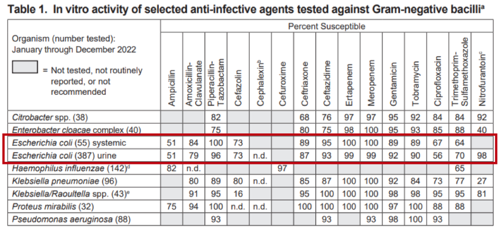
Shared Health Clinical Microbiology publishes hospital antibiogram data on a public website; the link is provided in our resources section, here. To see the antibiogram data for a given organism for the most recent year reported, we go to the website and select the year and relevant hospital. For example, if we want to look at the antibiogram data for E. coli at Seven Oaks Hospital (in 2023), we find the following antibiogram data (red box added for emphasis).

This means that among clinical urine (and blood) isolates tested, only 56% (and 67%) were susceptible to ciprofloxacin. This is not very good! And, while E. coli demonstrates high susceptibility rates to nitrofurantoin and fosfomycin (not shown in local antibiogram, because not enough isolates are tested by year, however national data including Manitoba reveals isolates are rarely resistant1), which may make these agents tempting to use for UTI, they are contraindicated in suspected pyelonephritis, such as in Bernadette’s case.

But there is more to consider when interpreting antibiogram data. READ ON!


Tips for Using Antibiograms in Practice

  • Antimicrobial resistance patterns vary by age, recent antibiotic treatment history, and underlying medical conditions for a given patient.
    • Always consider individual patient risk factors for antimicrobial resistance when interpreting antibiogram data.
    • A review of previous microbiology results (when available) can be very helpful.
  • Data from antibiograms may not be generalizable to specific populations.
    • For example, infections in otherwise healthy patients in the community are likely to have less antimicrobial resistance, than infections in hospitalized patients.
  • Beyond antimicrobial resistance, other important factors for clinicians to consider when selecting an empiric antimicrobial are:
    • the source of the infection and most likely pathogen(s) involved,
    • whether the infection is likely to be polymicrobial or monomicrobial,
    • the severity of the infection,
    • additional drug considerations, including side effects, oral absorption, and cost, and
    • additional patient factors including allergies, comorbidities, and potential drug-drug interactions.

Reference & Resource List:

1. Walkty A., Karlowsky JA et al. Microbiol Infect Dis. 2020 Mar; 96(3):114962. doi: 10.1016/j.diagmicrobio.2019.114962. Epub 2019 Dec 5.

The Canadian Antimicrobial Resistance Alliance (CARA) performs national surveillance of common bacterial pathogens and collates a national and regional (e.g. Western Canada) Antibiogram which can be useful to compare to Manitoba.

Interpreting the Data

It is important to appreciate that antibiograms are made up of clinical isolates submitted from hospitals, not from patients in the community, who tend to be healthier and exhibit less antibiotic resistance. Thus, data from antibiograms may not be generalizable to certain populations (See Limitations to Consider).  For example, bacterial infections occurring in the community (as in this case) are likely to have less antimicrobial resistance than infections in hospitalized patients.

For Bernadette, who is moderately ill with pyelonephritis, and has a history of recurrent UTIs (and presumably, multiple previous antibiotic exposures), it is advisable to choose an agent with >80-85% susceptibility. The goal is to choose an antibiotic likely to be effective, but to also consider patient-related factors, including individual risk factors for drug resistance and how ill the patient is (see Tips for Practice section, below). For Bernadette, empiric IV ceftriaxone or ceftazidime (with hospital-specific antibiogram susceptibility rates >80-85%) would be a better antibiotic choice while we give the clinical microbiology lab a chance to perform antimicrobial susceptibility testing on her E. coli. By the same token, if we were concerned that Bernadette was currently in septic shock (where the sepsis-driven mortality is dependent on time-to-effective antibiotic administration), the broader spectrum piperacillin-tazobactam or meropenem would be more appropriate (with a hospital-specific antibiogram susceptibility rate of 95% or higher).

As always, once the specific antimicrobial susceptibility rate of the E. coli is determined (which can take up to 36 hours in most cases), Bernadette’s antibiotics would be re-evaluated and changed to a narrower but equally effective agent, as appropriate.


IMPORTANT! – Limitations to Consider:

Correct interpretation of the information presented in hospital antibiograms requires an understanding of their limitations, as follows:

  • Hospital antibiograms are institution-specific.  It cannot be assumed that antimicrobial susceptibility rates at one healthcare facility will be identical to those at another. 
  • Susceptibility rates presented in hospital antibiograms for common bacterial pathogens should not be extrapolated to isolates obtained from specimens collected in the community (e.g., family physician’s office, outpatient microbiology laboratory). Community isolates are generally excluded from hospital antibiograms.
  • It is important to note that when antibiogram data are compiled, only the first isolate of a particular species from a patient is included over the defined time period. As a result, hospital antibiogram susceptibility rates are less helpful in guiding the selection of empiric therapy for patients who have received prior treatment with multiple antibiotic courses during a prolonged hospital stay. 
  • Hospital antibiograms do not contain data for all clinically relevant bacterial pathogens. For a bacterial pathogen to be included, there needs to be a minimum of 30 isolates tested on an annual basis.  

Case 10: Bronchiolitis – Christopher

Christopher is a 9-month-old girl who was brought into your local emergency room by her parents. She was born at term and has been healthy at her regular well-child visits. She has received all her recommended immunizations, including two-doses of influenza at six and seven months old, and her first Covid-19 immunization. Her parents describe a four-day history of cough and congestion. For the past two days she has had intermittent tactile fevers, and has been fussier. She is breastfed and eats various soft-solid foods. She has been eating less but is breastfeeding well and has been having four to five wet diapers each day. Both her parents and her two-year-old brother have had cough and rhinorrhea.

At triage, her vital signs are as follows: temperature is 38.5° Celsius, heart rate is 160 beats/min, respiratory rate is 50 breaths/min, oxygen saturation 94% on room air, and blood pressure is 93/58. She has been given a dose of acetaminophen prior to your assessment. On examination, you observe a settled infant with mildly increased work of breathing. There is slight subcostal indrawing, but no intercostal indrawing or nasal flaring. On auscultation, there are coarse crackles and scattered wheezes bilaterally. The remainder of her examination is unremarkable.

Question:

You suspect a diagnosis of bronchiolitis. What is the most appropriate next step?

  1. Order a chest x-ray to evaluate for pneumonia
  2. Provide nasal suction as needed and observe oral feeding in the emergency department
  3. Prescribe amoxicillin for treatment of community-acquired pneumonia
  4. Order a nasopharyngeal swab for QUADRUPLEX NAAT to evaluate for Influenza, RSV, and Covid-19
  5. Initiate continuous pulse oximetry and observe in the emergency department

Best response:

2.         Provide nasal suction as needed and observe oral feeding in the emergency department

Teaching Points:

Bronchiolitis is a viral infection of the lower respiratory tract occurring most commonly in infants between 1 and 12 months old. The pathophysiology of bronchiolitis includes necrosis of the respiratory epithelium and destruction of the ciliated layer, mononuclear dell invasion of the peribronchial tissues and alveolar interstitium, edema of the submucosa and adventitia, and small airway obstruction by plugs made of dead epithelial cells, fibrin, and inflammatory cells. Clinically, this presents with tachypnea, fever, cough, coryza, increased work of breathing, respiratory crackles, expiratory wheezing, and air trapping.

Bronchiolitis is caused by viral pathogens, most often respiratory syncytial virus (RSV), but less commonly due to parainfluenza viruses, rhinoviruses, metapneumovirus, adenoviruses, influenza viruses, enteroviruses, and Coronaviruses. It affects greater than 1/3 of all children before 2 years of life and is the most common cause of hospital admission for infants in Canada.

The diagnosis of bronchiolitis is based on history and physical examination.  The Canadian Paediatric Society position statement1 on bronchiolitis outlines key history and physical findings, as well as other considerations for a differential diagnosis. They also outline an algorithm for emergency room management, adapted from the American Academy of Pediatrics2. Choosing Wisely Canada3 and Trekk4 also outline evidence-based management recommendations for this common condition.

A routine chest radiograph is not required for the diagnosis or management of bronchiolitis. Findings are non-specific and can be misinterpreted as consolidation, leading to inappropriate use of antibiotics. A chest x-ray should be obtained when the diagnosis of bronchiolitis is unclear, or the severity of illness or rate of improvement suggests another diagnostic possibility, such as bacterial pneumonia. Continuous pulse oximetry is also not recommended for paediatric patients unless they require supplemental oxygen; transient desaturation may be captured, which in absence of other signs of respiratory distress are of uncertain clinical significance and may result in unnecessary oxygen administration and hospital admission. Nasopharyngeal swabs are also not required as the result will not change the disease management for the patient. An exception here is when nasaopharyngeal swab identification of viral pathogens may be useful for epidemiology or infection control surveillance, such as is the case in children who require hospitalization for bronchiolitis and may require cohorting with other patients with the same virus1,2,3

Antimicrobial therapy should not be initiated here. The management of bronchiolitis is supportive and focuses on providing adequate oxygenation and hydration. Nasal suction and epinephrine nebulization may be helpful in children requiring admission for bronchiolitis. The evidence for clinical benefit in all children presenting with bronchiolitis is equivocal, and neither intervention is routinely recommended for children not requiring admission1.

Children who do not meet admission criteria may be discharged home. Parents should be counselled on what to watch for and when to bring their child back for reassessment. Choosing Wisely Canada has published a “Viral Prescription” useful in assisting families understand viral infections and can be provided at the time of discharge5.


Admission Criteria for Bronchiolitis1

  • Signs of severe respiratory distress (severe work of breathing, persistent vital sign abnormalities)
  • Supplemental oxygen required to maintain SpO2 > 90%
  • Dehydration or poor oral fluid intake (often due to tachypnea)
  • Cyanosis or history of apnea
  • Infants at high risk for severe disease (infants born at less than 35 weeks gestation), very young infants (less than three months old), hemodynamically significant cardiopulmonary disease, or immunodeficiency
  • Caregivers unable to manage child at home

Resources and References: